Protein structures: Difference between revisions
GuentertWiki (talk | contribs) No edit summary |
GuentertWiki (talk | contribs) No edit summary |
||
| (36 intermediate revisions by the same user not shown) | |||
| Line 2: | Line 2: | ||
{| | {| | ||
|[[image:9wps.pdb-500.jpg|120px|link=http://www.rcsb.org/pdb/explore/explore.do?structureId=9WPS]] | |||
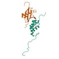
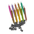
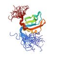
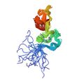
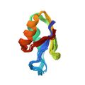
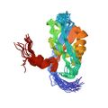
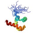
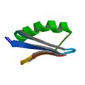
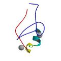
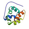
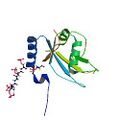
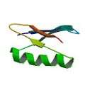
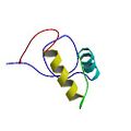
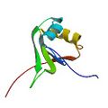
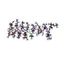
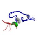
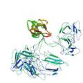
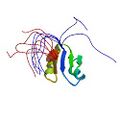
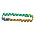
|Nameki, N., He, F., Okada, M., Takahashi, M., Tsuda, K., Nagata, T., Güntert, P., Kobayashi, N., Kigawa, T., Shirouzu, M., Tanaka, A., Yokoyama, S., Muto, Y., Kuwasako, K. Solution structure of mouse HBS1L/SKI7-specific UBA domain in complex with ubiquitin: Implications for stalled ribosome recognition[https://uni-frankfurt.guentert.science/Reprints/Nameki26-HBS1L.pdf .] [https://doi.org/10.1371/journal.pone.0348877 PLoS One 21, e0348877 (2026)] | |||
'''PDB [http://www.rcsb.org/pdb/explore.do?structureId=9WPR 9WPR]''' [http://files.rcsb.org/download/9wpr_nmr-data.nef.gz NMR data] '''BMRB [http://bmrb.io/data_library/generate_summary.php?bmrbId=36786 36786]''' (N-terminal domain of mouse HBS1L)<br> | |||
'''PDB [http://www.rcsb.org/pdb/explore.do?structureId=9WPS 9WPS]''' [http://files.rcsb.org/download/9wps_nmr-data.nef.gz NMR data] '''BMRB [http://bmrb.io/data_library/generate_summary.php?bmrbId=36787 36787]''' (complex between the UBA-like domain of mouse HBS1L and ubiquitin) | |||
|- | |||
|[[image:8ox2.pdb-500.jpg|120px|link=http://www.rcsb.org/pdb/explore/explore.do?structureId=8OX2]] | |[[image:8ox2.pdb-500.jpg|120px|link=http://www.rcsb.org/pdb/explore/explore.do?structureId=8OX2]] | ||
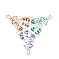
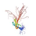
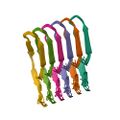
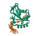
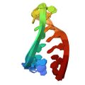
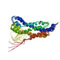
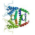
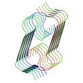
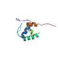
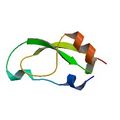
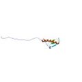
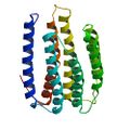
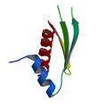
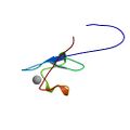
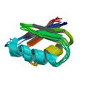
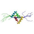
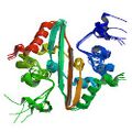
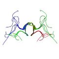
|Ghosh, D., Torres, F., Schneider, M. M., Ashkinadze, D., Kadavath, H., Fleischmann, Y., Mergenthal, S., Güntert, P. , Krainer, G., Andrzejewska, E. A., Lin, L., Wei, J., Klotzsch, E., Knowles, T. & Riek, R. The inhibitory action of the chaperone BRICHOS against α-Synuclein secondary nucleation pathway at near-atomic resolution[https://uni-frankfurt.guentert.science/Reprints/Ghosh24-BRICHOS.pdf .] [https://doi.org/10.1038/s41467-024-54212-2 Nat. Commun. 15, 10038 (2024)] | |Ghosh, D., Torres, F., Schneider, M. M., Ashkinadze, D., Kadavath, H., Fleischmann, Y., Mergenthal, S., Güntert, P. , Krainer, G., Andrzejewska, E. A., Lin, L., Wei, J., Klotzsch, E., Knowles, T. & Riek, R. The inhibitory action of the chaperone BRICHOS against α-Synuclein secondary nucleation pathway at near-atomic resolution[https://uni-frankfurt.guentert.science/Reprints/Ghosh24-BRICHOS.pdf .] [https://doi.org/10.1038/s41467-024-54212-2 Nat. Commun. 15, 10038 (2024)] | ||
'''PDB [http://www.rcsb.org/pdb/explore.do?structureId=8OVI 8OVI]''' [ | '''PDB [http://www.rcsb.org/pdb/explore.do?structureId=8OVI 8OVI]''' [http://files.rcsb.org/download/8ovi_nmr-data.nef.gz NMR data] '''BMRB [http://bmrb.io/data_library/generate_summary.php?bmrbId=34811 34811]''' (monomer)<br> | ||
'''PDB [http://www.rcsb.org/pdb/explore.do?structureId= | '''PDB [http://www.rcsb.org/pdb/explore.do?structureId=8OX2 8OX2]''' [http://files.rcsb.org/download/8ox2_nmr-data.nef.gz NMR data] '''BMRB [http://bmrb.io/data_library/generate_summary.php?bmrbId=34813 34813]''' (trimer) | ||
''' | |||
|- | |- | ||
|[[image:8kcq.pdb-500.jpg|120px|link=http://www.rcsb.org/pdb/explore/explore.do?structureId=8KCQ]] | |||
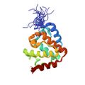
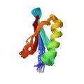
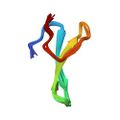
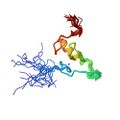
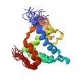
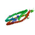
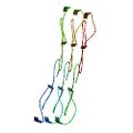
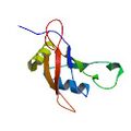
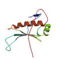
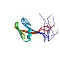
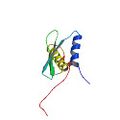
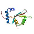
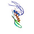
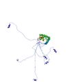
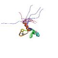
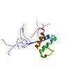
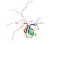
|Kuwasako, K., Dang, W., Fahu, H., Takahashi, M., Tsuda, K., Nagata, T., Tanaka, A., Kobayashi, N., Kigawa, T., Güntert, P., Shirouzu, M., Yokoyama, S. & Muto, Y. <sup>1</sup>H, <sup>13</sup>C and <sup>15</sup>N resonance assignments and solution structure of the N-terminal divergent caplonin homology (NN-CH) domain of human intraflagellar transport protein 54[https://uni-frankfurt.guentert.science/Reprints/Kuwasako24-Protein54Assignment.pdf .] [http://doi.org/10.1007/s12104-024-10170-w Biomol. NMR Assignments 18, 71–78 (2024)] | |||
'''PDB [http://www.rcsb.org/pdb/explore.do?structureId=8KCQ 8KCQ]''' [http://files.rcsb.org/download/8ox2_nmr-data.nef.gz NMR data] '''BMRB [http://bmrb.io/data_library/generate_summary.php?bmrbId=36589 36589]''' | |||
|- | |||
|[[image:8qbp.pdb-500.jpg|120px|link=http://www.rcsb.org/pdb/explore/explore.do?structureId=8QBP]] | |||
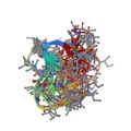
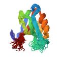
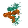
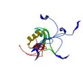
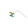
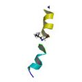
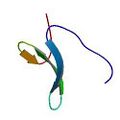
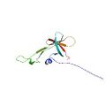
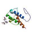
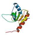
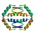
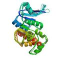
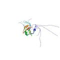
|Rüdisser, S. H., Matabaro, E., Sonderegger, L., Güntert, P., Künzler, M., Gossert, A. D. Conformations of macrocyclic peptides sampled by exact NOEs: Models for cell-permeability[https://uni-frankfurt.guentert.science/Reprints/Ruedisser23-eNOEPeptides.pdf .] [http://doi.org/10.1021/jacs.3c09367 J. Am. Chem. Soc. 145, 27601–27615 (2023)] | |||
'''PDB [http://www.rcsb.org/pdb/explore.do?structureId=8QBP 8QBP]''' [http://files.rcsb.org/download/8qbp_nmr-data.nef.gz NMR data] '''BMRB [https://bmrb.io/data_library/generate_summary.php?bmrbId=34850 34850]''' (OmphA “indole-In) | |||
<br> | |||
'''PDB [http://www.rcsb.org/pdb/explore.do?structureId=8Q7J 8Q7J]''' [http://files.rcsb.org/download/8q7j_nmr-data.nef.gz NMR data] '''BMRB [https://bmrb.io/data_library/generate_summary.php?bmrbId=34846 34846]''' (CsA) | |||
<br> | |||
'''PDB [http://www.rcsb.org/pdb/explore.do?structureId=8QAQ 8QAQ]''' [http://files.rcsb.org/download/8qaq_nmr-data.nef.gz NMR data] '''BMRB [https://bmrb.io/data_library/generate_summary.php?bmrbId=34848 34848]''' (OmphA | |||
conformation 1) | |||
<br> | |||
'''PDB [http://www.rcsb.org/pdb/explore.do?structureId=8QAS 8QAS]''' [http://files.rcsb.org/download/8qas_nmr-data.nef.gz NMR data] '''BMRB [https://bmrb.io/data_library/generate_summary.php?bmrbId=34849 34849]''' (OmphA “indole-Out”) | |||
|- | |||
|[[image:8pxs.pdb-500.jpg|120px|link=http://www.rcsb.org/pdb/explore/explore.do?structureId=8PXS]] | |||
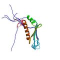
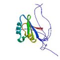
|Rout, S. K., Cadalbert, R., Schröder, N., Wang, J., Zehnder, J., Gampp, O., Wiegand, T., Güntert, P. , Klingler, D., Kreutz, C., Knörlein, A., Hall, J., Greenwald, J. & Riek, R. An analysis of nucleotide-amyloid interactions reveals selective binding to codon-sized RNA[https://uni-frankfurt.guentert.science/Reprints/Rout23-RNAmyloid.pdf .] [https://doi.org/10.1021/jacs.3c06287 J. Am. Chem. Soc. 145, 21915−21924 (2023)] | |||
'''PDB [http://www.rcsb.org/pdb/explore.do?structureId=8PXS 8PXS]''' [http://files.rcsb.org/download/8pxs_nmr-data.nef.gz NMR data] '''BMRB [https://bmrb.io/data_library/generate_summary.php?bmrbId=34838 34838]''' | |||
|- | |||
|[[image:7x9u.pdb-500.jpg|120px|link=http://www.rcsb.org/pdb/explore/explore.do?structureId=7X9U]] | |||
|Kuwasako, K., Suzuki, S., Nameki, N., Takizawa, M., Takahashi, M., Tsuda, K., Nagata, T., Watanabe, S., Tanaka, A., Kobayashi, N., Kigawa, T., Güntert, P., Shirouzu, M., Yokoyama, S. & Muto, Y. <sup>1</sup>H, <sup>13</sup>C and <sup>15</sup>N resonance assignments and solution structures of the KH domain of ribosome binding factor A, mtRbfA, from humans, that is involved in mitochondrial ribosome biogenesis[https://uni-frankfurt.guentert.science/Reprints/Kuwasako22-mtRbfA.pdf .] [http://doi.org/10.1007/s12104-022-10094-3 Biomol. NMR Assignments 16, 297–303 (2022)] | |||
'''PDB [http://www.rcsb.org/pdb/explore.do?structureId=7X9U 7X9U]''' [http://files.rcsb.org/download/7x9u_nmr-data.nef.gz NMR data] '''BMRB [https://bmrb.io/data_library/generate_summary.php?bmrbId=36478 36478]''' | |||
|- | |||
|[[image:7qtr.pdb-500.jpg|120px|link=http://www.rcsb.org/pdb/explore/explore.do?structureId=7QTR]] | |||
|Gerez, J. A., Prymaczok, N. C., Kadavath, H., Ghosh, D., Bütikofer, M., Güntert, P. & Riek, R. Protein structure determination in human cells by in-cell NMR and a reporter system to optimize protein delivery or transexpression[https://uni-frankfurt.guentert.science/Reprints/Gerez22-HumanInCellNMR.pdf .] [http://doi.org/10.1038/s42003-022-04251-6 Comm. Biol. 5, 1322 (2022)] | |||
'''PDB [http://www.rcsb.org/pdb/explore.do?structureId=7QTR 7QTR]''' [http://files.rcsb.org/download/7qtr_nmr-data.nef.gz NMR data] '''BMRB [https://bmrb.io/data_library/generate_summary.php?bmrbId=34700 34700]''' (GB1 in cells at a protein concentration of 50 μM) | |||
<br> | |||
'''PDB [http://www.rcsb.org/pdb/explore.do?structureId=7QTS 7QTS]''' [http://files.rcsb.org/download/7qts_nmr-data.nef.gz NMR data] '''BMRB [https://bmrb.io/data_library/generate_summary.php?bmrbId=34701 34701]''' (GB1 in cells at a protein concentration of 10 μM) | |||
|- | |||
|[[image:7qcx.pdb-500.jpg|120px|link=http://www.rcsb.org/pdb/explore/explore.do?structureId=7QCX]] | |||
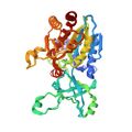
|Ashkinadze, D., Kadavath, H., Chi, C., Friedmann, M., Strotz, D., Kumari, P., Minges, M., Cadalbert, C., Königl, S., Güntert, P., Vögeli, B. & Riek, R. Atomic resolution protein allostery from the multi-state structure of a PDZ Domain[https://uni-frankfurt.guentert.science/Reprints/Ashkinadze22-PDZ.pdf .] [http://doi.org/10.1038/s41467-022-33687-x Nat. Commun. 13, 6232 (2022)] | |||
'''PDB [http://www.rcsb.org/pdb/explore.do?structureId=7QCX 7QCX]''' [http://files.rcsb.org/download/7qcx_nmr-data.nef.gz NMR data] '''BMRB [https://bmrb.io/data_library/generate_summary.php?bmrbId=34688 34688]''' (two-state structure of apo PDZ2) | |||
<br> | |||
'''PDB [http://www.rcsb.org/pdb/explore.do?structureId=7QCY 7QCY]''' [http://files.rcsb.org/download/7qcy_nmr-data.nef.gz NMR data] '''BMRB [https://bmrb.io/data_library/generate_summary.php?bmrbId=34689 34689]''' (two-state structure of PDZ2 complexed with RA-GEF2 peptide) | |||
|- | |||
|[[image:7fbr.pdb-500.jpg|120px|link=http://www.rcsb.org/pdb/explore/explore.do?structureId=7FBR]] | |||
|He., F., Kuwasako, K., Takizawa, M., Takahashi, M., Tsuda, K., Nagata, T., Watanabe, S., Tanaka, A., Kobayashi, N., Kigawa, T., Güntert, P., Shirouzu, M., Yokoyama, S. & Muto, Y. <sup>1</sup>H, <sup>13</sup>C and <sup>15</sup>N resonance assignments and solution structures of the two RRM domains of Matrin-3[https://uni-frankfurt.guentert.science/Reprints/He22-Matrin3.pdf .] [http://doi.org/10.1007/s12104-021-10057-0 Biomol. NMR Assignments 16, 41–49 (2022)] | |||
'''PDB [http://www.rcsb.org/pdb/explore.do?structureId=7FBR 7FBR]''' [http://files.rcsb.org/download/7fbr_nmr-data.nef.gz NMR data] '''BMRB [https://bmrb.io/data_library/generate_summary.php?bmrbId=36430 36430]''' (first RRM domain of Matrin-3) | |||
<br> | |||
'''PDB [http://www.rcsb.org/pdb/explore.do?structureId=7FBV 7FBV]''' [http://files.rcsb.org/download/7fbv_nmr-data.nef.gz NMR data] '''BMRB [https://bmrb.io/data_library/generate_summary.php?bmrbId=36431 36431]''' (second RRM domain of Matrin-3) | |||
|- | |||
|[[image:6tub.pdb-500.jpg|120px|link=http://www.rcsb.org/pdb/explore/explore.do?structureId=6TUB]] | |||
|Seuring, C., Verasdonck, J., Gath, J., Ghosh, D., Nespovitaya, N., Wälti, M. A., Maji, S., Cadalbert, R., Güntert, P., Meier, B. H. & Riek, R. The three-dimensional structure of human β-endorphin amyloid fibrils[https://uni-frankfurt.guentert.science/Reprints/Seuring20-Endorphin.pdf .] [http://doi.org/10.1038/s41594-020-00515-z Nat. Struct. Mol. Biol. 27, 1178–1184 (2020)] | |||
'''PDB [http://www.rcsb.org/pdb/explore.do?structureId=6TUB 6TUB]''' [http://files.rcsb.org/download/6tub_nmr-data.nef.gz NMR data] '''BMRB [https://bmrb.io/data_library/generate_summary.php?bmrbId=26715 26715]''' | |||
|- | |||
|[[image:6svc.pdb-500.jpg|120px|link=http://www.rcsb.org/pdb/explore/explore.do?structureId=6SVE]] | |||
| Strotz, D., Orts, J., Kadavath, H., Friedmann, M., Ghosh, D., Olsson, S., Chi, C. N., Pokharna, A., Güntert, P., Vögeli, B. & Riek, R. Protein allostery at atomic resolution[https://uni-frankfurt.guentert.science/Reprints/Strotz20-WWAllostery.pdf .] [http://doi.org/10.1002/anie.202008734 Angew. Chem. Int. Ed. 59, 22132-22139 (2020)] | |||
'''PDB [http://www.rcsb.org/pdb/explore.do?structureId=6SVC 6SVC]''' [http://files.rcsb.org/download/6svc_nmr-data.nef.gz NMR data] '''BMRB [https://bmrb.io/data_library/generate_summary.php?bmrbId=34432 34432]''' | |||
(WW domain apo structure) | |||
<br> | |||
'''PDB [http://www.rcsb.org/pdb/explore.do?structureId=6SVE 6SVE]''' [http://files.rcsb.org/download/6sve_nmr-data.nef.gz NMR data] '''BMRB [https://bmrb.io/data_library/generate_summary.php?bmrbId=34433 34433]''' (WW domain with pCdc25C bound) | |||
<br> | |||
'''PDB [http://www.rcsb.org/pdb/explore.do?structureId=6SVH 6SVH]''' [http://files.rcsb.org/download/6svh_nmr-data.nef.gz NMR data] '''BMRB [https://bmrb.io/data_library/generate_summary.php?bmrbId=34434 34434]''' (WW domain with FFpSPR bound) | |||
|- | |||
|[[image:6qk6.pdb-500.jpg|120px|link=http://www.rcsb.org/pdb/explore/explore.do?structureId=6QK6]] | |||
|Beil, A., Jurt, S., Walser, R., Schäfer, T., Güntert, P., Capdeville, M., Atrian, S., Dallinger, R. & Zerbe, O. The solution structure and dynamics of Cd-metallothionein from ''Helix pomatia'' reveal optimization for binding Cd over Zn. Biochemistry[https://uni-frankfurt.guentert.science/Reprints/Beil19-HpMT.pdf .] [http://doi.org/10.1021/acs.biochem.9b00830 Biochemistry 58, 4570-4581 (2019)] | |||
'''PDB [http://www.rcsb.org/pdb/explore.do?structureId=6QK5 6QK5]''' [http://files.rcsb.org/download/6qk5_nmr-data.nef.gz NMR data] '''BMRB [https://bmrb.io/data_library/generate_summary.php?bmrbId=34355 34355]''' (Zn-loaded form) | |||
<br> | |||
'''PDB [http://www.rcsb.org/pdb/explore.do?structureId=6QK6 6QK6]''' [http://files.rcsb.org/download/6qk6_nmr-data.nef.gz NMR data] '''BMRB [https://bmrb.io/data_library/generate_summary.php?bmrbId=34356 34356]''' (Cd-loaded form) | |||
|- | |||
|[[image:5zcz.pdb-500.jpg|120px|link=http://www.rcsb.org/pdb/explore/explore.do?structureId=5ZCZ]] | |||
|Tanaka, T., Ikeya, T., Kamoshida, H., Suemoto, Y., Mishima, M., Shirakawa, M., Güntert, P., Ito, Y. High resolution protein 3D structure determination in living eukaryotic cells[https://uni-frankfurt.guentert.science/Reprints/Tanaka19-EukaryoticInCellNMR.pdf .] [http://doi.org/10.1002/anie.201900840 Angew. Chem. Int. Ed. 58, 7284–7288 (2019)] | |||
'''PDB [http://www.rcsb.org/pdb/explore.do?structureId=5ZCZ 5ZCZ]''' [http://files.rcsb.org/download/5zcz_nmr-data.nef.gz NMR data] '''BMRB [https://bmrb.io/data_library/generate_summary.php?bmrbId=27354 27354]''' (''T. thermophilus'' HB8 TTHA1718 protein in living eukaryotic cells) | |||
<br> | |||
'''PDB [http://www.rcsb.org/pdb/explore.do?structureId=5ZD0 5ZD0]''' [http://files.rcsb.org/download/5zd0_nmr-data.nef.gz NMR data] '''BMRB [https://bmrb.io/data_library/generate_summary.php?bmrbId=27356 27356]''' (ubiquitin with three alanine mutations in living eukaryotic cells) | |||
<br> | |||
'''PDB [http://www.rcsb.org/pdb/explore.do?structureId=5Z4B 5Z4B]''' [http://files.rcsb.org/download/5z4b_nmr-data.nef.gz NMR data] '''BMRB [https://bmrb.io/data_library/generate_summary.php?bmrbId=36153 36153]''' (GB1 in living eukaryotic cells) | |||
|- | |||
|[[image:6h8c.pdb-500.jpg|120px|link=http://www.rcsb.org/pdb/explore/explore.do?structureId=6H8C]] | |||
|Huber, J., Obata, M., Gruber, J., Akutsu, M., Löhr, F., Rogova, N., Güntert, P., Dikic, I., Krikin, V., Komatsu, M., Dötsch, V. & Rogov, V. V. An atypical LIR motif within UBA5 (ubiquitin like modifier activating enzyme 5) interacts with GABARAP proteins and mediates membrane localization of UBA5[https://uni-frankfurt.guentert.science/Reprints/Huber20-UBA5.pdf .] [http://doi.org/10.1080/15548627.2019.1606637 Autophagy 16, 256-270 (2020)] | |||
'''PDB [http://www.rcsb.org/pdb/explore.do?structureId=6H8C 6H8C]''' [http://files.rcsb.org/download/6h8c_nmr-data.nef.gz NMR data] '''BMRB [https://bmrb.io/data_library/generate_summary.php?bmrbId=34307 34307]''' (GABARAPL2 in complex with UBA5 LIR motif) | |||
<br> | |||
'''PDB [http://www.rcsb.org/pdb/explore.do?structureId=6HB9 6HB9]''' [http://files.rcsb.org/download/6hb9_nmr-data.nef.gz NMR data] (crystal structure of GABARAP in complex with UBA5 LIR motif) | |||
|- | |||
|[[image:6f98.pdb-500.jpg|120px|link=http://www.rcsb.org/pdb/explore/explore.do?structureId=6F98]] | |||
|Kniss, A., Kazemi, S., Löhr, F., Berger, M., Rogov, V. V., Güntert, P., Sommer, T., Jarosch, E. & Dötsch, V. Structural investigation of glycan recognition by the ERAD quality control lectin Yos9[https://uni-frankfurt.guentert.science/Reprints/Kniss18-Yos9.pdf .] [http://doi.org/10.1007/s10858-018-0201-6 J. Biomol. NMR 72, 1-10 (2018)] | |||
'''PDB [http://www.rcsb.org/pdb/explore.do?structureId=6F98 6F98]''' [http://files.rcsb.org/download/6f98_nmr-data.nef.gz NMR data] '''BMRB [https://bmrb.io/data_library/generate_summary.php?bmrbId=34217 34217]''' (RING domain of E3 ubiquitin ligase HRD1) | |||
<br> | |||
'''PDB [http://www.rcsb.org/pdb/explore.do?structureId=6F99 6F99]''' [http://files.rcsb.org/download/6f99_nmr-data.nef.gz NMR data] '''BMRB [https://bmrb.io/data_library/generate_summary.php?bmrbId=34218 34218]''' (MRH domain of Yos9) | |||
<br> | |||
'''PDB [http://www.rcsb.org/pdb/explore.do?structureId=6F9A 6F9A]''' [http://files.rcsb.org/download/6f9a_nmr-data.nef.gz NMR data] '''BMRB [https://bmrb.io/data_library/generate_summary.php?bmrbId=34219 34219]''' (MRH domain of Yos9 complexed with alpha3,alpha6-Man5) | |||
|- | |||
|[[image:6es5.pdb-500.jpg|120px|link=http://www.rcsb.org/pdb/explore/explore.do?structureId=6ES5]] | |||
|Jemth, P., Andersson, E., Vögeli, B., Hultqvist, G., Dogan, J., Güntert, P. & Riek, R. & Chi, C. N., Structure and dynamics conspire in the evolution of affinity between intrinsically disordered proteins[https://uni-frankfurt.guentert.science/Reprints/Jemth18-IDPEvolution.pdf .] [http://doi.org/10.1126/sciadv.aau4130 Science Adv. 4, eaau4130 (2018)] | |||
'''PDB [http://www.rcsb.org/pdb/explore.do?structureId=6ES5 6ES5]''' [http://files.rcsb.org/download/6es5_nmr-data.nef.gz NMR data] '''BMRB [https://bmrb.io/data_library/generate_summary.php?bmrbId=34187 34187]''' | |||
<br> | |||
'''PDB [http://www.rcsb.org/pdb/explore.do?structureId=6ES6 6ES6]''' [http://files.rcsb.org/download/6es6_nmr-data.nef.gz NMR data] '''BMRB [https://bmrb.io/data_library/generate_summary.php?bmrbId=34188 34188]''' | |||
<br> | |||
'''PDB [http://www.rcsb.org/pdb/explore.do?structureId=6ES7 6ES7]''' [http://files.rcsb.org/download/6es7_nmr-data.nef.gz NMR data] '''BMRB [https://bmrb.io/data_library/generate_summary.php?bmrbId=34189 34189]''' | |||
|- | |||
|[[image:6esp.pdb-500.jpg|120px|link=http://www.rcsb.org/pdb/explore/explore.do?structureId=6ESP]] | |||
|Sundell, G. N., Arnold, R., Ali, M., Naksukpaiboon, P., Orts, J., Güntert, P., Chi, C. N., Ivarsson, Y. Proteome-wide analysis of phospho-regulated PDZ domain interactions[https://uni-frankfurt.guentert.science/Reprints/Sundell18-PDZ.pdf .] [http://doi.org/10.15252/msb.20178129 Mol. Sys. Biol. 14, e8129 (2018)] | |||
'''PDB [http://www.rcsb.org/pdb/explore.do?structureId=6ESP 6ESP]''' [http://files.rcsb.org/download/6esp_nmr-data.nef.gz NMR data] '''BMRB [https://bmrb.io/data_library/generate_summary.php?bmrbId=34190 34190]''' | |||
|- | |||
|[[image:6by4.pdb-500.jpg|120px|link=http://www.rcsb.org/pdb/explore/explore.do?structureId=6BY4]] | |||
|Nichols, P.J., Henen, M.A., Born, A., Strotz, D., Guntert, P., Vogeli, B. <br> High-resolution small RNA structures from exact nuclear Overhauser enhancement measurements without additional restraints. [https://doi.org/10.1038/s42003-018-0067-x .] [Commun Biol 2018] | |||
'''PDB [http://www.rcsb.org/pdb/explore.do?structureId=6BY4 6BY4]''' [http://files.rcsb.org/download/6by4_nmr-data.nef.gz NMR data] '''BMRB [https://bmrb.io/data_library/generate_summary.php?bmrbId=30386 30386]''' | |||
<br> | |||
'''PDB [http://www.rcsb.org/pdb/explore.do?structureId=6BY5 6BY5]''' [http://files.rcsb.org/download/6by5_nmr-data.nef.gz NMR data] '''BMRB [https://bmrb.io/data_library/generate_summary.php?bmrbId=30386 30386]''' | |||
|- | |||
|[[image:6fgn.pdb-500.jpg|120px|link=http://www.rcsb.org/pdb/explore/explore.do?structureId=6FGN]] | |||
|Krauskopf, K., Gebel, J., Kazemi, S., Löhr, F., Tuppi, M., Löhr, F., Schäfer, B., Koch, J., Güntert, P., Dötsch, V. & Kehrloesser, S. Regulation of the activity in the p53 family depends on the organization of the transactivation domain[https://uni-frankfurt.guentert.science/Reprints/Krauskopf18-p53TAD.pdf .] [http://doi.org/10.1016/j.str.2018.05.013 Structure 26, 1091–1100 (2018)] | |||
'''PDB [http://www.rcsb.org/pdb/explore.do?structureId=6FGN 6FGN]''' [http://files.rcsb.org/download/6fgn_nmr-data.nef.gz NMR data] '''BMRB [https://bmrb.io/data_library/generate_summary.php?bmrbId=34231 34231]''' | |||
<br> | |||
'''PDB [http://www.rcsb.org/pdb/explore.do?structureId=6FGS 6FGS]''' [http://files.rcsb.org/download/6fgs_nmr-data.nef.gz NMR data] '''BMRB [https://bmrb.io/data_library/generate_summary.php?bmrbId=34233 34233]''' | |||
|- | |||
|[[image:6f3k.pdb-500.jpg|120px|link=http://www.rcsb.org/pdb/explore/explore.do?structureId=6F3K]] | |||
|Gauto, D. F., Estrozi, L. F., Schwieters, C. D., Effantin, G., Macek, P., Sounier, R., Sivertsen, A. C., Schmidt, E., Kerfah, R., Mas, G., Colletier, J.-P., Güntert, P., Favier, A., Schoehn, G., Schanda, P. & Boisbouvier, J. Integrated NMR and cryo-EM atomic-resolution structure determination of a half-megadalton enzyme complex[https://uni-frankfurt.guentert.science/Reprints/Gauto19-TET2.pdf .] [http://doi.org/10.1038/s41467-019-10490-9 Nat. Commun. 10, 2697 (2019)] | |||
'''PDB [http://www.rcsb.org/pdb/explore.do?structureId=6F3K 6F3K]''' [http://files.rcsb.org/download/6f3k_nmr-data.nef.gz NMR data] '''BMRB [https://bmrb.io/data_library/generate_summary.php?bmrbId=27211 27211]''' (NMR + EM structure) | |||
<br> | |||
'''PDB [http://www.rcsb.org/pdb/explore.do?structureId=6R8N 6R8N]''' [http://files.rcsb.org/download/6r8n_nmr-data.nef.gz NMR data] '''BMRB [https://bmrb.io/data_library/generate_summary.php?bmrbId=27211 27211]''' (NMR + EM structure after Phenix refinement) | |||
|- | |||
|[[image:5n2o.pdb-500.jpg|120px|link=http://www.rcsb.org/pdb/explore/explore.do?structureId=5N2O]] | |||
|Russo, C., Osterburg, C., Sirico, A., Antonini, D., Ambrosio, R., Würz, J. M., Rinnental J., Schäfer, B., Güntert, P., Sinha, S., Dötsch, V. & Missero, C. Protein aggregation of the p63 transcription factor underlies severe skin fragility in AEC syndrome[https://uni-frankfurt.guentert.science/Reprints/Russo18-p63Aggregation.pdf .] [http://doi.org/10.1073/pnas.1713773115 Proc. Natl. Acad. Sci. USA 115, E906-E915 (2018)] | |||
'''PDB [http://www.rcsb.org/pdb/explore.do?structureId=5N2O 5N2O]''' [http://files.rcsb.org/download/5n2o_nmr-data.nef.gz NMR data] '''BMRB [https://bmrb.io/data_library/generate_summary.php?bmrbId=34095 34095]''' | |||
|- | |||
|[[image:5n6r.pdb-500.jpg|120px|link=http://www.rcsb.org/pdb/explore/explore.do?structureId=5N6R]] | |[[image:5n6r.pdb-500.jpg|120px|link=http://www.rcsb.org/pdb/explore/explore.do?structureId=5N6R]] | ||
Latest revision as of 06:50, 4 June 2026
NMR protein structures co-authored by P. Güntert.
|
Nameki, N., He, F., Okada, M., Takahashi, M., Tsuda, K., Nagata, T., Güntert, P., Kobayashi, N., Kigawa, T., Shirouzu, M., Tanaka, A., Yokoyama, S., Muto, Y., Kuwasako, K. Solution structure of mouse HBS1L/SKI7-specific UBA domain in complex with ubiquitin: Implications for stalled ribosome recognition. PLoS One 21, e0348877 (2026)
PDB 9WPR NMR data BMRB 36786 (N-terminal domain of mouse HBS1L) |
|
Ghosh, D., Torres, F., Schneider, M. M., Ashkinadze, D., Kadavath, H., Fleischmann, Y., Mergenthal, S., Güntert, P. , Krainer, G., Andrzejewska, E. A., Lin, L., Wei, J., Klotzsch, E., Knowles, T. & Riek, R. The inhibitory action of the chaperone BRICHOS against α-Synuclein secondary nucleation pathway at near-atomic resolution. Nat. Commun. 15, 10038 (2024)
PDB 8OVI NMR data BMRB 34811 (monomer) |
|
Kuwasako, K., Dang, W., Fahu, H., Takahashi, M., Tsuda, K., Nagata, T., Tanaka, A., Kobayashi, N., Kigawa, T., Güntert, P., Shirouzu, M., Yokoyama, S. & Muto, Y. 1H, 13C and 15N resonance assignments and solution structure of the N-terminal divergent caplonin homology (NN-CH) domain of human intraflagellar transport protein 54. Biomol. NMR Assignments 18, 71–78 (2024) |
|
Rüdisser, S. H., Matabaro, E., Sonderegger, L., Güntert, P., Künzler, M., Gossert, A. D. Conformations of macrocyclic peptides sampled by exact NOEs: Models for cell-permeability. J. Am. Chem. Soc. 145, 27601–27615 (2023)
PDB 8QBP NMR data BMRB 34850 (OmphA “indole-In)
|
|
Rout, S. K., Cadalbert, R., Schröder, N., Wang, J., Zehnder, J., Gampp, O., Wiegand, T., Güntert, P. , Klingler, D., Kreutz, C., Knörlein, A., Hall, J., Greenwald, J. & Riek, R. An analysis of nucleotide-amyloid interactions reveals selective binding to codon-sized RNA. J. Am. Chem. Soc. 145, 21915−21924 (2023) |
|
Kuwasako, K., Suzuki, S., Nameki, N., Takizawa, M., Takahashi, M., Tsuda, K., Nagata, T., Watanabe, S., Tanaka, A., Kobayashi, N., Kigawa, T., Güntert, P., Shirouzu, M., Yokoyama, S. & Muto, Y. 1H, 13C and 15N resonance assignments and solution structures of the KH domain of ribosome binding factor A, mtRbfA, from humans, that is involved in mitochondrial ribosome biogenesis. Biomol. NMR Assignments 16, 297–303 (2022) |
|
Gerez, J. A., Prymaczok, N. C., Kadavath, H., Ghosh, D., Bütikofer, M., Güntert, P. & Riek, R. Protein structure determination in human cells by in-cell NMR and a reporter system to optimize protein delivery or transexpression. Comm. Biol. 5, 1322 (2022)
PDB 7QTR NMR data BMRB 34700 (GB1 in cells at a protein concentration of 50 μM)
|
|
Ashkinadze, D., Kadavath, H., Chi, C., Friedmann, M., Strotz, D., Kumari, P., Minges, M., Cadalbert, C., Königl, S., Güntert, P., Vögeli, B. & Riek, R. Atomic resolution protein allostery from the multi-state structure of a PDZ Domain. Nat. Commun. 13, 6232 (2022)
PDB 7QCX NMR data BMRB 34688 (two-state structure of apo PDZ2)
|
|
He., F., Kuwasako, K., Takizawa, M., Takahashi, M., Tsuda, K., Nagata, T., Watanabe, S., Tanaka, A., Kobayashi, N., Kigawa, T., Güntert, P., Shirouzu, M., Yokoyama, S. & Muto, Y. 1H, 13C and 15N resonance assignments and solution structures of the two RRM domains of Matrin-3. Biomol. NMR Assignments 16, 41–49 (2022)
PDB 7FBR NMR data BMRB 36430 (first RRM domain of Matrin-3)
|
|
Seuring, C., Verasdonck, J., Gath, J., Ghosh, D., Nespovitaya, N., Wälti, M. A., Maji, S., Cadalbert, R., Güntert, P., Meier, B. H. & Riek, R. The three-dimensional structure of human β-endorphin amyloid fibrils. Nat. Struct. Mol. Biol. 27, 1178–1184 (2020) |
|
Strotz, D., Orts, J., Kadavath, H., Friedmann, M., Ghosh, D., Olsson, S., Chi, C. N., Pokharna, A., Güntert, P., Vögeli, B. & Riek, R. Protein allostery at atomic resolution. Angew. Chem. Int. Ed. 59, 22132-22139 (2020)
PDB 6SVC NMR data BMRB 34432
(WW domain apo structure)
|
|
Beil, A., Jurt, S., Walser, R., Schäfer, T., Güntert, P., Capdeville, M., Atrian, S., Dallinger, R. & Zerbe, O. The solution structure and dynamics of Cd-metallothionein from Helix pomatia reveal optimization for binding Cd over Zn. Biochemistry. Biochemistry 58, 4570-4581 (2019)
PDB 6QK5 NMR data BMRB 34355 (Zn-loaded form)
|
|
Tanaka, T., Ikeya, T., Kamoshida, H., Suemoto, Y., Mishima, M., Shirakawa, M., Güntert, P., Ito, Y. High resolution protein 3D structure determination in living eukaryotic cells. Angew. Chem. Int. Ed. 58, 7284–7288 (2019)
PDB 5ZCZ NMR data BMRB 27354 (T. thermophilus HB8 TTHA1718 protein in living eukaryotic cells)
|
|
Huber, J., Obata, M., Gruber, J., Akutsu, M., Löhr, F., Rogova, N., Güntert, P., Dikic, I., Krikin, V., Komatsu, M., Dötsch, V. & Rogov, V. V. An atypical LIR motif within UBA5 (ubiquitin like modifier activating enzyme 5) interacts with GABARAP proteins and mediates membrane localization of UBA5. Autophagy 16, 256-270 (2020)
PDB 6H8C NMR data BMRB 34307 (GABARAPL2 in complex with UBA5 LIR motif)
|
|
Kniss, A., Kazemi, S., Löhr, F., Berger, M., Rogov, V. V., Güntert, P., Sommer, T., Jarosch, E. & Dötsch, V. Structural investigation of glycan recognition by the ERAD quality control lectin Yos9. J. Biomol. NMR 72, 1-10 (2018)
PDB 6F98 NMR data BMRB 34217 (RING domain of E3 ubiquitin ligase HRD1)
|
|
Jemth, P., Andersson, E., Vögeli, B., Hultqvist, G., Dogan, J., Güntert, P. & Riek, R. & Chi, C. N., Structure and dynamics conspire in the evolution of affinity between intrinsically disordered proteins. Science Adv. 4, eaau4130 (2018)
PDB 6ES5 NMR data BMRB 34187
|
|
Sundell, G. N., Arnold, R., Ali, M., Naksukpaiboon, P., Orts, J., Güntert, P., Chi, C. N., Ivarsson, Y. Proteome-wide analysis of phospho-regulated PDZ domain interactions. Mol. Sys. Biol. 14, e8129 (2018) |
|
Nichols, P.J., Henen, M.A., Born, A., Strotz, D., Guntert, P., Vogeli, B. High-resolution small RNA structures from exact nuclear Overhauser enhancement measurements without additional restraints. . [Commun Biol 2018] |
|
Krauskopf, K., Gebel, J., Kazemi, S., Löhr, F., Tuppi, M., Löhr, F., Schäfer, B., Koch, J., Güntert, P., Dötsch, V. & Kehrloesser, S. Regulation of the activity in the p53 family depends on the organization of the transactivation domain. Structure 26, 1091–1100 (2018) |
|
Gauto, D. F., Estrozi, L. F., Schwieters, C. D., Effantin, G., Macek, P., Sounier, R., Sivertsen, A. C., Schmidt, E., Kerfah, R., Mas, G., Colletier, J.-P., Güntert, P., Favier, A., Schoehn, G., Schanda, P. & Boisbouvier, J. Integrated NMR and cryo-EM atomic-resolution structure determination of a half-megadalton enzyme complex. Nat. Commun. 10, 2697 (2019)
PDB 6F3K NMR data BMRB 27211 (NMR + EM structure)
|
|
Russo, C., Osterburg, C., Sirico, A., Antonini, D., Ambrosio, R., Würz, J. M., Rinnental J., Schäfer, B., Güntert, P., Sinha, S., Dötsch, V. & Missero, C. Protein aggregation of the p63 transcription factor underlies severe skin fragility in AEC syndrome. Proc. Natl. Acad. Sci. USA 115, E906-E915 (2018) |
|
Reckel, S., Gehin, C., Tardivon, D., Harduin, D., Georgeon, S., Kükenshöner, T., Löhr, F., Koide, A., Buchner, L., Panjkovich, A., Reynaud, A., Pinho, S., Gerig, B., Svergun, D., Pojer, F., Güntert, P., Dötsch, V., Koide, S., Gavin, A.-C. & Hantschel, O. Structural and functional dissection of the DH and PH domains of oncogenic Bcr-Abl tyrosine kinase. Nat. Commun. 8, 2101 (2017)
PDB 5N6R NMR restraints BMRB 34101 (DH NMR structure) |
|
Bibow, S., Polyhach, Y., Eichmann, C., Chi, C. N., Kowal, J., Albiez, S., McLeod, R. A., Stahlberg, H., Jeschke, G., Güntert, P. & Riek, R. Solution structure of discoidal high-density lipoprotein particles with a shortened apolipoprotein A-I. Nat. Struct. Mol. Biol. 24, 187-193 (2017)
PDB 2N5E NMR restraints BMRB 25710 |
|
Kuwasako, K., Nameki, N., Tsuda, K., Takahashi, M., Sato, A., Tochio, N., Inoue, M., Terada, T., Kigawa, T., Kobayashi, N., Shirouzu, M., Ito, T., Sakamoto, T., Wakamatsu, K., Güntert, P., Takahashi, S., Yokoyama, S. & Muto, Y. Solution structure of the first RNA recognition motif domain of human spliceosomal protein SF3b49 and its mode of interaction with a SF3b145 fragment. Protein Sci. 26, 280-291 (2017)
PDB 5GVQ NMR restraints BMRB 36018 |
|
Ikeya, T., Hanashima, T., Hosoya, S., Shimazaki, M., Ikeda, S., Mishima, M., Güntert, P. & Ito, Y. Improved in-cell structure determination of proteins at near-physiological concentration. Sci Rep. 6, 38312 (2016)
PDB 2N9L NMR restraints BMRB 25910 |
|
Gebel, J., Luh, L. M., Coutandin, D., Osterburg, C., Löhr, F., Schäfer, B., Frombach, A., Sumyk, M., Buchner, L., Krojer, T., Salah, E., Mathea, S., Güntert, P., Knapp, S. & Dötsch, V. Mechanism of TAp73 inhibition by ΔNp63 and structural basis of p63/p73 hetero-tetramerization. Cell Death Diff. 23, 1930-1940 (2016)
PDB 2NB1 NMR restraints BMRB 25958 |
|
Wälti, M. A., Ravotti, F., Arai, H., Glabe, C., Wall, J., Böckmann, A., Güntert, P., Meier, B. H. & Riek, R. Atomic resolution structure of a disease-relevant Aβ(1–42) amyloid fibril. Proc. Natl. Acad. Sci. USA
PDB 2NAO NMR restraints BMRB 26692 |
|
von Delbrück, M., Kniss, A., Rogov, V. V., Pluska, L., Bagola, K., Löhr, F., Güntert, P., Sommer, T. & Dötsch, V. The CUE domain of Cue1 aligns growing ubiquitin chains with Ubc7 for rapid elongation. Mol. Cell 62, 918-928 (2016)
PDB 2MYX NMR restraints BMRB 25461 |
|
Huang, S. Y., Chang, C. F., Fan, P. J., Naik, M. T., Güntert, P., Shih, H. M. & Huang, T. H. The RING domain of human promyelocytic leukemia protein (PML). J. Biomol. NMR 61, 173-180 (2015)
PDB 2MWX NMR restraints BMRB 25376 |
|
Schütz, A. K., Vagt, T., Huber, M., Ovchinnikova, O. Y., Cadalbert, R., Wall, J., Güntert, P., Böckmann, A., Glockshuber, R., and Meier, B. H. Atomic-resolution three-dimensional structure of amyloid β fibrils bearing the Osaka mutation. Angew. Chem. Int. Ed. 54, 331–335 (2015)
PDB 2MVX NMR restraints BMRB 25289 |
|
Tsuda, K., Kuwasako, K., Nagata, T., Takahashi, M., Kigawa, T., Kobayashi, N., Güntert, P., Shirouzu, M., Yokoyama, S. & Muto, Y. Novel RNA recognition motif domain in cytoplasmic polyadenylation element binding protein 3. Proteins 82, 2879–2886 (2014)
PDB 2RUG NMR restraints BMRB 11563 |

|
Kuwasako, K., Takahashi, M., Unzai, S., Tsuda, K., Yoshikawa, S., He, F., Kobayashi, N., Güntert, P., Shirouzu, M., Ito, T., Tanaka, A., Yokoyama, S., Hagiwara, M., Kuroyanagi, H. & Muto, Y. RBFOX and SUP-12 sandwich a guanine base to cooperatively regulate tissue-specific splicing. Nat. Struct. Mol. Biol. 21, 778–786 (2014)
PDB 2RU3 NMR restraints BMRB 11517 (SUP-12–RNA6 complex) |
|
Uggerhøj, L. E., Munk, J. K., Hansen, P. R., Güntert, P. & Wimmer, R. Structural features of peptoid-peptide hybrids in lipid-water interfaces. FEBS Lett. 588, 3291–3297 (2014)
PDB 2MMJ NMR restraints BMRB 19856 (M-Nleu11) |
|
Watson, R. P., Christen, M. T., Bumbak, F., Ewald, C., Reichen, C. Mihajlovic, M., Schmidt, E., Güntert, P., Caflisch, A., Plückthun, A., Zerbe, O. Spontaneous self assembly of fragments of engineered Armadillo repeat proteins into a folded structure. Structure 22, 985–995 (2014)
PDB 2RU5 NMR restraints BMRB 11548 (uncomplexed MA) |
|
Tufar, P., Rahighi, S., Kraas, F. I., Kirchner, D. K., Löhr, F., Henrich, E., Köpke, J., Dikic, I., Güntert, P., Marahiel, M. A. & Dötsch, V. Crystal structure of a PCP/Sfp complex reveals the structural basis for carrier protein posttranslational modification. Chem. Biol. 21, 552–562 (2014)
PDB 2MD9 NMR restraints BMRB 19479 |
|
Kogure, H., Handa, Y., Nagata, M., Kanai, N., Güntert, P., Kubota, K. & Nameki, N. Identification of residues required for stalled-ribosome rescue in the codon-independent release factor YaeJ. Nucl. Acids Res. 42, 3152-3163 (2014)
PDB 2RTX NMR restraints BMRB 11534 |
|
Luh, L. M., Hänsel, R., Löhr, F., Kirchner, D. K., Krauskopf, K., Pitzius, S., Schäfer, B., Tufar, P., Corbeski, I., Güntert, P. & Dötsch, V. Molecular crowding drives active Pin1 into nonspecific complexes with endogenous proteins prior to substrate recognition. J. Am. Chem. Soc. 135, 13796−13803 (2013)
PDB 2M8I NMR restraints BMRB 19258 (wildtype
WW domain) |
|
Rogov, V. V., Suzuki, H., Fiskin, E., Wild, P., Kniss, A., Rozenknop, A., Kato, R., Kawasaki, M., McEwan, D. G., Löhr, F., Güntert, P., Dikic, I., Wakatsuki, S. & Dötsch, V. Structural basis for phosphorylation-triggered autophagic clearance of Salmonella. Biochem. J. 454, 459–466 (2013)
PDB 2LUE NMR restraints BMRB 18518 (NMR structure of LC3B OPTN-LIR Ptot complex) |
|
Lin, Y. J., Ikeya, T., Güntert, P. & Chang, L. S. NMR solution structure of a chymotrypsin inhibitor from the Taiwan cobra. Molecules 18, 8906-8918 (2013)
PDB 2M99 NMR restraints BMRB 19287 |
|
He, F., Tsuda, K., Takahashi, M., Kuwasako, K., Terada, T., Shirouzu, M., Watanabe, S., Kigawa, T., Kobayashi, N., Güntert, P., Yokoyama, S. & Muto, Y. Structural insight into the interaction of ADP-ribose with the PARP WWE domains. FEBS Lett. 586, 3858–3864 (2012)
PDB 2DK6 NMR restraints BMRB 11500 (WWE domain from PARP11) |
|
Christ, N. A., Bochmann, S., Gottstein, D., Duchardt-Ferner, E., Hellmich, U. A., Düsterhus, S., Kötter, P., Güntert, P., Entian, K.-D. & Wöhnert, J. The first structure of a lantibiotic immunity protein, SpaI from Bacillus subtilis, reveals a novel fold. J. Biol. Chem. 287, 35286-35298 (2012)
PDB 2LVL NMR restraints BMRB 17534 |
|
Vögeli, B., Kazemi, S., Güntert, P. & Riek, R. Spatial elucidation of motion in proteins by ensemble-based structure calculation using exact NOEs. Nature Struct. Mol. Biol. 19, 1053–1057 (2012)
PDB 2LUM NMR restraints BMRB 18531 |
|
Kogure, H., Hikawa, Y., Hagihara, M., Tochio, N., Koshiba, S., Inoue, Y., Güntert, P., Kigawa, T., Yokoyama, S. & Nameki, N. Solution structure and siRNA-mediated knockdown analysis of the mitochondrial disease-related protein C12orf65. Proteins 80, 2629–2642 (2012)
PDB 2RSM NMR restraints BMRB 11491 |
|
Nagata, T., Tsuda, K., Shirouzu, M., Kigawa, T., Kobayashi, N., Güntert, P., Yokoyama, S. & Muto, Y. Solution structures of the double-stranded RNA-binding domains from RNA-helicase A. Proteins 80, 1699–1706 (2012)
PDB 2RS6 NMR restraints BMRB 11456 (dsRBD1) |
|
Busche, A., Gottstein, D., Hein, C., Ripin, N., Pader, I., Tufar, P., Eisman, E. B., Gu, L. Walsh, C. T., Sherman, D. H., Löhr, F., Güntert, P. & Dötsch, V. Characterization of the interaction between an ACP domain and a halogenase in the curacin A polyketide synthetase. ACS Chem. Biol. 7, 378–386 (2012)
PDB 2LIU NMR restraints BMRB 17906 (holo-ACPI) |
|
He, F., Inoue, M., Kigawa, T., Takahashi, M., Kuwasako, K., Tsuda, K., Kobayashi, N., Terada, T., Shirouzu, M., Tanaka, A., Sugano, S., Güntert, P., Yokoyama, S. & Muto, Y. Solution structure of the Splicing Factor Motif (SFM) of the human Prp18 protein. Proteins 80, 968–974 (2012)
PDB 2DK4 NMR restraints BMRB 11355 |
|
Reckel, S., Gottstein, D., Stehle, J., Löhr, F., Verhoefen, M. K., Takeda, M., Silvers, R., Kainosho, M., Glaubitz, C., Wachtveitl, J., Bernhard, F., Schwalbe, H., Güntert, P. & Dötsch, V. Solution NMR structure of proteorhodopsin. Angew. Chem. Int. Ed. 50, 11942–11946 (2011)
PDB 2L6X NMR restraints BMRB 17327 |
|
Rozenknop, A., Rogov, V. V., Löhr, F., Güntert, P., Dikic, I., & Dötsch, V. Characterization of the interaction of GABARAPL-1 with the LIR motif of NBR1. J. Mol. Biol. 410, 477–487 (2011)
PDB 2L8J NMR restraints BMRB 17412 |
|
Schmöe, K., Rogov, V. V., Rogova, N. Y., Löhr, F., Güntert, P., Bernhard, F. & Dötsch, V. Structural insights into Rcs phosphotransfer: The newly identified RcsD-ABL domain enhances interaction with the response regulator RcsB. Structure 19, 577–587 (2011)
PDB 2KX7 NMR restraints |
|
Tsuda, K., Someya, T., Kuwasako, K., Takahashi, M., Fahu, H., Inoue, M., Harada, T., Watanabe, S., Terada, T., Kobayashi, N., Shirouzu, M., Kigawa, T., Tanaka, A., Sugano, S., Güntert, P., Yokoyama, S. & Muto, Y. Structural basis for the dual RNA-recognition modes of human Tra2-β RRM. Nucl. Acids Res. 39, 1538-1553 (2011)
PDB 2RRB NMR restraints |
|
Yamashita, S., Nagata, T., Kawazoe, M., Takemoto, C., Kigawa, T, Güntert, P., Kobayashi, N., Terada, T., Shirouzu, M., Wakiyama, M., Muto, Y. & Yokoyama, S. Structures of the first and second double-stranded RNA-binding domains of human TAR RNA-binding protein. Protein Sci. 20, 118-130 (2011) |
|
Yamashita, S., Nagata, T., Kawazoe, M., Takemoto, C., Kigawa, T, Güntert, P., Kobayashi, N., Terada, T., Shirouzu, M., Wakiyama, M., Muto, Y. & Yokoyama, S. Structures of the first and second double-stranded RNA-binding domains of human TAR RNA-binding protein. Protein Sci. 20, 118-130 (2011)
PDB 2CPN NMR restraints |
|
Handa, Y., Hikawa, Y., Tochio, N., Kogure, H., Inoue, M., Koshiba, S., Güntert, P., Inoue, Y., Kigawa, T., Yokoyama, S. & Nameki, N. Solution structure of the mitochondrial protein ICT1 that is essential for cell vitality. J. Mol. Biol. 404, 260-273 (2010)
PDB 1J26 |
|
Hamada, T., Matsunaga, S.,Fujiwara, M., Fujita, K., Hirota, H., Schmucki, R., Güntert, P. & Fusetani, N. Solution structure of polytheonamide B, a highly cytotoxic nonribosomal polypeptide from marine sponge. J. Am. Chem. Soc. 132, 12941–12945 (2010)
PDB 2RQO NMR restraints |
|
He, F., Umehara, T., Saito, K., Harada, T., Watanabe, S., Yobuki, T., Kigawa, T., Takahashi, M., Kuwasako, K., Tsuda, K., Matsuda, T., Aoki, M., Seki, E., Kobayashi, N., Güntert, P., Yokoyama, S. & Muto, Y. Structural insight into the zinc finger CW domain as a histone modification reader. Structure 18, 1127–1139 (2010)
PDB 2E61 NMR restraints |
|
Sobhanifar, S., Schneider, B., Löhr, F., Gottstein, D., Ikeya, T., Filipek, S., Güntert, P., Bernhard, F. & Dötsch, V. Structural investigation of the C-terminal catalytic fragment of presenilin-1. Proc. Natl. Acad. Sci. USA 107, 9644–9649 (2010)
PDB 2KR6 NMR restraints |
|
Coutandin, D., Löhr, F., Niesen, F. H., Ikeya, T., Weber, T. A., Schäfer, B., Bullock, A. N., Yang, A., Güntert, P. , Knapp, S., McKeon, F., Der Ou, H. & Dötsch, V. Conformational stability and activity of p73 require a second helix in the tetramerization domain. Cell Death Diff. 16, 1582–1589 (2009)
PDB 2KBY NMR restraints |
|
He, F., Saito, K., Kobayashi, N., Harada, T., Watanabe, S., Kigawa, T, Güntert, P., Unzai, S., Muto, Y. & Yokoyama, S. Structural and functional characterization of the NHR1 domain of the Drosophila Neuralized E3 ligase in the Notch signaling pathway. J. Mol. Biol. 393, 478-495 (2009)
PDB 2YUE NMR restraints |
|
Khayrutdinov, B. I., Bae, W. J., Yun, Y. M., Lee, J. H., Tsuyama, T., Kim, J. J., Hwang, E., Ryu, K. S., Cheong, H. K., Cheong, C., Ko, J. S., Enomoto, T., Karplus, P. A., Güntert, P., Tada, S., Jeon, Y. H., Cho, Y. Structure of the Cdt1 C-terminal domain: Conservation of the winged helix fold in replication licensing factors. Protein Sci. 18, 2252-2264 (2009)
PDB 2KLO NMR restraints |
|
Tsuda, K., Kuwasako, K., Takahashi, M., Someya, T., Inoue, M., Terada, T., Kobayashi, N., Shirouzu, M., Kigawa, T., Tanaka, A., Sugano, S., Güntert, P., Muto, Y. & Yokoyama, S. Structural basis for the sequence specific RNA-recognition mechanism of human CUG-BP1 RRM3. Nucl. Acids Res. 37, 5151–5166 (2009)
PDB 2RQC NMR restraints |
|
He, F., Dang, W., Saito, K., Watanabe, S., Kobayashi, N., Güntert, P., Kigawa, T, Tanaka, A., Muto, Y. & Yokoyama, S. Solution structure of the cysteine-rich domain in Fn14, a member of the tumor necrosis factor receptor superfamily. Protein Sci. 18, 650–656 (2009)
PDB 2RPJ NMR restraints |
|
Peroza, E. A., Schmucki, R., Güntert, P., Freisinger, E. & Zerbe O. The βE-domain of Wheat Ec-1 metallothionein: A metal-binding domain with a distinctive structure. J. Mol. Biol. 387, 207–218 (2009)
PDB 2KAK NMR restraints BMRB 16025 |
|
Sakakibara, D., Sasaki, A., Ikeya, T., Hamatsu, J., Hanashima, T., Mishima, M., Yoshimasu, M., Hayashi, N., Mikawa, T., Wälchli, M., Smith, B. O., Shirakawa, M., Güntert, P. & Ito, Y. Protein structure determination in living cells by in-cell NMR spectroscopy. Nature 458, 102-105 (2009)
PDB 2ROG NMR restraints BMRB 11037 (in-cell) |
|
He, F., Dang, W., Abe, C., Tsuda, K., Inoue, M., Watanabe, S., Kobayashi, N., Kigawa, T, Matsuda, T., Yabuki, T., Aoki, M., Seki, E., Harada, T., Tomabechi, Y., Terada, T., Shirouzu, M., Tanaka, A., Güntert, P., Muto, Y. & Yokoyama, S. Solution structure of the RNA binding domain in the human muscleblind-like protein 2. Protein Sci. 18, 80-91 (2009)
PDB 2E5S NMR restraints |
|
Ohnishi, S. Pääkkönen, K., Koshiba, S., Tochio, N., Sato, M., Kobayashi, N., Harada, T., Watanabe, S., Muto, Y., Güntert, P., Tanaka, A., Kigawa, T. & Yokoyama, S. Solution structure of the GUCT domain from human RNA helicase II/Guβ reveals the RRM fold, but implausible RNA interactions. Proteins 74, 133–144 (2009)
PDB 2E29 |
|
Takeda, M., Sugimori, N., Torizawa, T., Terauchi, T., Ono, A. M., Yagi, H., Yamaguchi, Y., Kato, K., Ikeya, T., Jee, J., Güntert, P., Aceti, D. J., Markley, J. L. & Kainosho, M. Structure of the putative 32 kDa myrosinase binding protein from Arabidopsis (At3g16450.1) as determined by the SAIL-NMR method. FEBS J. 275, 5873–5884 (2008)
PDB 2JZ4 NMR restraints BMRB 15607 |
|
Yoshida, H., Furuya, N., Lin, Y. J., Güntert, P., Komano, T. & Kainosho, M. Structural basis of the role of the NikA ribbon-helix-helix domain in initiating bacterial conjugation. J. Mol. Biol. 384, 690–701 (2008) |
|
Lin, Y. J., Umehara, T., Inoue, M., Saito, K., Kigawa, T., Jang, M. K., Ozato, K., Yokoyama, S., Padmanabhan, B., Güntert, P. Solution structure of the extraterminal domain of the bromodomain-containing protein BRD4. Protein Sci. 17, 2174–2179 (2008) |
|
Koglin, A., Löhr, F., Bernhard, F., Rogov, V. R., Frueh, D. P., Strieter, E. R., Mofid, M. R., Güntert, P., Wagner, G., Walsh, C. T., Marahiel, M. A. & Dötsch, V. Structural basis for the selectivity of the external thioesterase of the surfactin-synthetase. Nature 454, 907–911 (2008)
PDB 2RON NMR restraints (SrfTEII) |
|
Nagata, T., Suzuki, S., Endo, R., Shirouzu, M., Terada, T., Inoue, M., Kigawa, T, Güntert, P., Hayashizaki, Y., Muto, Y. & Yokoyama, S. The RRM domain of poly(A)-specific ribonuclease (PARN) has a non-canonical binding site for mRNA cap analog recognition. Nucl. Acids Res. 36, 4754–4767 (2008) |
|
Takeda, M., Chang, C. K., Ikeya, T., Güntert, P., Chang, Y. H., Hsu, Y. L., Huang, T. H. & Kainosho, M. Solution structure of the C-terminal dimerization domain of SARS coronavirus nucleocapsid protein determined by the SAIL-NMR method. J. Mol. Biol. 380, 608–622 (2008) |
|
Kuwasako, K., Takahashi, M., Tochio, N., Abe, C., Tsuda, K., Inoue, M., Terada, T., Shirouzu, M., Kobayashi, N., Kigawa, T., Taguchi, S., Tanaka, A., Hayashizaki, Y., Güntert, P., Muto, Y. & Yokoyama, S. Solution structure of the second RNA recognition motif (RRM) domain of murine T cell intracellular antigen-1 (TIA-1) and its RNA recognition mode. Biochemistry 47, 6437–6450 (2008)
PDB 2RNE |
|
Kuwasako, K., Dohmae, N., Inoue, M., Shirouzu, M., Taguchi, S., Güntert, P., Séraphin, B., Muto, Y. & Yokoyama, S. Complex assembly mechanism and an RNA-binding mode of the human p14-SF3b155 spliceosomal protein complex identified by NMR solution structure and functional analyses. Proteins 71, 1617–1636 (2008)
PDB 2FHO NMR restraints |
|
Hwang, E., Ryu, K. S., Pääkkönen, K., Güntert, P., Cheong, H. K., Lim, D. S., Lee, J. O., Jeon, Y. H. & Cheong, C. Structural insight into dimeric interaction of the SARAH domains from Mst1 and RASSF family proteins in the apoptosis pathway. Proc. Natl. Acad. Sci. USA. 104, 9236–9241 (2007)
PDB 2JO8 NMR restraints |
|
Ohnishi, S. Güntert, P., Koshiba, S., Tomizawa, T., Akasaka, R., Tochio, N., Sato, M., Inoue, M., Harada, T., Watanabe, S., Tanaka, T., Shirouzu, M., Kigawa, T. & Yokoyama, S. Solution structure of an atypical WW domain in a novel β-clam-like dimeric form. FEBS Lett. 581, 462–468 (2007)
PDB 2DWV |

|
Kuwasako, K., He, F., Inoue, M., Tanaka, A., Sugano, S., Güntert, P., Muto, Y. & Yokoyama, S. Solution structures of the SURP domains and the subunit-assembly mechanism within the splicing factor SF3a complex in 17S U2 snRNP. Structure 14, 1677–1689 (2006) |

|
ENTH-VHS domain At3g16270(9-135) from Arabidopsis thaliana.
López-Méndez, B. & Güntert, P. Automated protein structure determination from NMR spectra. J. Am. Chem. Soc. 128, 13112–13122 (2006) PDB 2DCP NMR restraints Note: Automated structure determination with FLYA. Structure does not supersede the original deposition 1VDY. |

|
Rhodanese homology domain At3g16270(175-295) from Arabidopsis thaliana.
López-Méndez, B. & Güntert, P. Automated protein structure determination from NMR spectra. J. Am. Chem. Soc. 128, 13112–13122 (2006) PDB 2DCQ NMR restraints Note: Automated structure determination with FLYA. Structure does not supersede the original deposition 1VEE. |

|
Src homology domain 2 from the human feline sarcoma oncogene Fes.
López-Méndez, B. & Güntert, P. Automated protein structure determination from NMR spectra. J. Am. Chem. Soc. 128, 13112–13122 (2006) PDB 2DCR NMR restraintsbr>Note: Automated structure determination with FLYA. Structure does not supersede the original deposition 1WQU. |

|
Jurt, S., Aemissegger, A., Güntert, P., Zerbe, O. & Hilvert, D. A photoswitchable miniprotein based on the sequence of avian pancreatic polypeptide. Angew. Chem. Int. Ed. 45, 6297-6300 (2006)
PDB 2H4B NMR restraints (cis-1-PP) |

|
Hamada, T., Ito, Y., Abe, T., Hayashi, F., Güntert, P., Inoue, M., Kigawa, T., Terada, T., Shirouzu, M., Yoshida, M., Tanaka, A., Sugano, S., Yokoyama, S. & Hirota, H. Solution structure of the antifreeze-like domain of human sialic acid synthase. Protein Sci. 15, 1010–1016 (2006)
PDB 1WVO NMR restraints |

|
Aachmann, F. L., Svanem, B. I. G., Güntert, P., Petersen, S. B., Valla, S. & Wimmer, R. NMR structure of the R-module - A parallel β-roll subunit from an Azotobacter vinelandii mannuronan C-5 epimerase. J. Biol. Chem. 281, 7350–7356 (2006)
PDB 2AGM NMR restraints |

|
Stereo-array isotope labeled (SAIL) maltodextrin-binding protein MBP.
Kainosho, M., Torizawa, T., Iwashita, Y., Terauchi, T., Ono, A. M. & Güntert, P. Optimal isotope labeling for NMR protein structure determinations. Nature 440, 52–57 (2006) |

|
Stereo-array isotope labeled (SAIL) calmodulin.
Kainosho, M., Torizawa, T., Iwashita, Y., Terauchi, T., Ono, A. M. & Güntert, P. Optimal isotope labeling for NMR protein structure determinations. Nature 440, 52–57 (2006) |

|
Li, H., Inoue, M., Yabuki, T., Aoki, M., Seki, E., Matsuda, T., Nunokawa, E., Motoda, Y., Kobayashi, A., Terada, T., Shirouzu, M., Koshiba, S., Lin, Y. J., Güntert, P., Suzuki, H., Hayashizaki, Y., Kigawa, T. & Yokoyama, S. Solution structure of the mouse enhancer of rudimentary protein reveals a novel fold. J. Biomol. NMR 32, 329–334 (2005)
PDB 1WWQ |

|
Scott, A., Pantoja-Uceda, D., Koshiba, S., Inoue, M., Kigawa, T., Terada, T., Shirouzu, M., Tanaka, A., Sugano, S., Yokoyama, S. & Güntert, P. Solution structure of the Src homology 2 domain from the human feline sarcoma oncogene Fes. J. Biomol. NMR. 31, 357–361 (2005) |

|
Nameki, N., Tochio, N., Koshiba, S., Inoue, M., Yabuki, T., Aoki, M., Seki, E., Matsuda, T., Fujikura, Y., Saito, M., Ikari, M., Watanabe, M., Terada, T., Shirouzu, M., Yoshida, M., Hirota, H., Tanaka, A., Hayashizaki, Y., Güntert, P., Kigawa, T. & Yokoyama, S. Solution structure of the PWWP domain of the hepatoma-derived growth factor family. Protein Sci. 14, 756–764 (2005)
PDB 1N27 |

|
Calzolai, L., Lysek, D. A., Pérez, D. R., Güntert, P. & Wüthrich, K. Prion protein NMR structures of chicken, turtle and frog. Proc. Natl. Acad. Sci. USA 102, 651–655 (2005)
PDB 1U3M NMR restraints BMRB 6269 (chicken PrP(121-225)) |

|
Lysek, D. A., Schorn, C., Nivon, L. G., Esteve-Moya, V., Christen, B., Calzolai, L., von Schroetter, C., Fiorito, F., Herrmann, T., Güntert, P. & Wüthrich, K. Prion protein NMR structures of cat, dog, pig and sheep. Proc. Natl. Acad. Sci. USA 102, 640–645 (2005)
PDB 1XYJ NMR restraints BMRB 6377 (cat PrP(121-231)) |

|
Pääkkönen, K., Tossavainen, H., Permi, P., Rakkolainen, H., Rauvala, H., Raulo, E., Kilpeläinen, I. & Güntert, P. Solution structures of the first and fourth TSR domains of F-spondin. Proteins 64, 665–672 (2006)
PDB 1SZL NMR restraints BMRB 6175 (TSR1) |

|
ENTH-VHS domain At3g16270 from Arabidopsis thaliana. |

|
Iwai, H., Forrer, P., Plückthun, A. & Güntert, P. NMR solution structure of the monomeric form of the bacteriophage λ capsid stabilizing protein gpD. J. Biomol. NMR. 31, 351–356 (2005) |

|
Pantoja-Uceda, D., López-Méndez, B., Koshiba, S., Inoue, M., Kigawa, T., Terada, T., Shirouzu, M., Tanaka, A., Seki, M., Shinozaki, K., Yokoyama, S. & Güntert, P. Solution structure of the rhodanese homology domain At4g01050(175–295) from Arabidopsis thaliana. Protein Sci. 14, 224–230 (2005) |

|
Nameki, N., Yoneyama, M., Koshiba, S., Tochio, N., Inoue, M., Seki, E., Matsuda, T., Tomo, Y., Harada, T., Saito, K., Kobayashi, N., Yabuki, T., Aoki, M., Nunokawa, E., Matsuda, N., Sakagami, N., Terada, T., Shirouzu, M., Yoshida, M., Hirota, H., Osanai, T., Tanaka, A., Arakawa, T., Carninci, P., Kawai, J., Hayashizaki, Y., Kinoshita, K., Güntert, P., Kigawa, T. & Yokoyama, S. Solution structure of the RWD domain of the mouse GCN2 protein. Protein Sci. 13, 2089–2100 (2004) |

|
Fernández, C., Hilty, C., Wider, G., Güntert, P. & Wüthrich, K. NMR structure of the integral membrane protein OmpX. J. Mol. Biol. 336, 1211–1221 (2004) |

|
FHA domain of mouse afadin 6.
PDB 1WLN |

|
Vanwetswinkel, S., Kriek, J., Andersen, G. R., Güntert, P., Dijk, P., Canters, G. W. & Siegal, G. Solution structure of the 162 residue C-terminal domain of human elongation factor 1Bγ. J. Biol. Chem. 278, 43443–43451 (2003)
PDB 1PBU NMR restraints BMRB 5628 |

|
Hilge, M., Siegal, G., Vuister, G. W., Güntert, P., Gloor, S. M. & Abrahams, J. P. ATP-induced conformational changes of the nucleotide-binding domain of Na,K-ATPase. Nat. Struct. Biol. 10, 468–474 (2003)
PDB 1MO7 BMRB 5577 (free form) |

|
Lührs, T., Riek, R., Güntert, P. & Wüthrich, K. NMR structure of the human doppel protein. J. Mol. Biol. 326, 1549–1557 (2003)
PDB 1LG4 NMR restraints BMRB 5145 |

|
Zahn, R., Güntert, P. & Wüthrich, K. NMR structure of a variant human prion protein with two disulfide bonds. J. Mol. Biol. 326, 225–234 (2003)
PDB 1H0L NMR restraints BMRB 5378 |

|
Enggist, E., Thöny-Meyer, L., Güntert, P. & Pervushin, K. NMR structure of the heme chaperone CcmE reveals a new functional motif. Structure 10, 1551–1557 (2002)
PDB 1SR3 NMR restraints |

|
Lee, D., Damberger, F. D., Peng, G., Horst, R., Güntert, P., Nikonova, L., Leal, W. S. & Wüthrich, K. NMR structure of the unliganded Bombyx mori pheromone-binding protein at physiological pH. FEBS Lett. 531, 314–318 (2002)
PDB 1LS8 |

|
Ellgaard, L., Bettendorff, P., Braun, D., Herrmann, T., Fiorito, F., Jelesarov, I., Güntert, P., Helenius, A. & Wüthrich, K. NMR Structures of 36 and 73-residue fragments of the calreticulin P-domain. J. Mol. Biol. 322, 773–784 (2002)
PDB 1K9C NMR restraints BMRB 5204 (residues 189-261) |

|
Miura, T., Klaus, W., Ross, A., Güntert, P., Senn, H. The NMR structure of the class I human ubiquitin-conjugating enzyme 2b. J. Biomol. NMR 22, 89–92 (2002)
PDB 1JAS NMR restraints BMRB 5038 |

|
Horst, R., Damberger, F., Luginbühl, P., Güntert, P., Peng, G., Nikonova, L., Leal, W. S. & Wüthrich, K. NMR structure reveals intramolecular regulation mechanism for pheromone binding and release. Proc. Natl. Acad. Sci. USA 98, 14374–14379 (2001)
PDB 1GM0 NMR restraints BMRB 4849 |

|
Ellgaard, L., Riek, R., Herrmann, T., Güntert, P., Braun, D., Helenius, A. & Wüthrich, K. NMR structure of the calreticulin P-domain. Proc. Natl. Acad. Sci. USA 98, 3133–3138 (2001) |

|
Calzolai, L., Lysek, D. A., Güntert, P., von Schroetter, C., Riek, R., Zahn, R. & Wüthrich, K. NMR structures of three single-residue variants of the human prion protein. Proc. Natl. Acad. Sci. USA 97, 8340–8345 (2000)
PDB 1E1G NMR restraints BMRB 4736 (variant M166V) |

|
Riek, R., Prêcheur, B., Wang, Y., Wider, G., Güntert, P., Liu, A., Kägi, J. H. R. & Wüthrich, K. NMR structure of the sea urchin (Strongylocentrotus purpuratus) metallothionein MTA. J. Mol. Biol. 291, 417–428 (1999)
PDB 1QJK NMR restraints (α domain) |

|
Pellecchia, M., Güntert, P., Glockshuber, R. & Wüthrich, K. The NMR solution structure of the periplasmic chaperone FimC. Nature Struct. Biol. 5, 885–890 (1998)
PDB 1BF8 NMR restraints BMRB 4070 |

|
Ottiger, M., Zerbe, O., Güntert, P. & Wüthrich, K. The NMR solution conformation of unligated human Cyclophilin A. J. Mol. Biol. 272, 64–81 (1997)
PDB 1OCA NMR restraints |

|
Antuch, W., Güntert, P. & Wüthrich, K. Ancestral βγ-crystallin precursor structure in a yeast killer toxin. Nature Struct. Biol. 3, 662–665 (1996)
PDB 1WKT NMR restraints BMRB 5255 |

|
Antuch, W., Güntert, P., Billeter, M., Hawthorne, T., Grossenbacher, H. & Wüthrich, K. NMR solution structure of the recombinant tick anticoagulant protein (rTAP), a factor Xa inhibitor from the tick Ornithodoros moubata. FEBS Lett. 352, 251–257 (1994)
PDB 1TAP NMR restraints |

|
Berndt, K. D., Güntert, P. & Wüthrich, K. The NMR solution structure of dendrotoxin K from the venom of Dendroaspis polylepis polylepis. J. Mol. Biol. 234, 735–750 (1993)
PDB 1DTK NMR restraints |

|
Szyperski, T., Güntert, P., Stone, S. R. & Wüthrich, K. NMR solution structure of hirudin(1–51) and comparison with corresponding three-dimensional structures determined using the complete 65-residue hirudin polypeptide chain. J. Mol. Biol. 228, 1193–1205 (1992)
PDB 1HIC NMR restraintsBMRB 1128 |

|
Berndt, K. D., Güntert, P., Orbons, L. P. M. & Wüthrich, K. Determination of a high-quality NMR solution structure of the bovine pancreatic trypsin inhibitor (BPTI) and comparison with three crystal structures. J. Mol. Biol. 227, 757–775 (1992)
PDB 1PIT NMR restraints |

|
Güntert, P., Qian, Y. Q., Otting, G., Müller, M., Gehring, W. J. & Wüthrich K. Structure determination of the Antp(C39S) homeodomain from nuclear magnetic resonance data in solution using a novel strategy for the structure calculation with the programs DIANA, CALIBA, HABAS and GLOMSA. J. Mol. Biol. 217, 531–540 (1991)
PDB 2HOA |